LDI
Moderated by Ir. Bas Generowicz, PhD-student Department of Neuroscience, Erasmus MC, Rotterdam
Laser Doppler Imaging (LDI) is a doppler-based optical technique, which makes use of the fact that laser light scatters on moving particles, leading to a frequency shifted signal (the Doppler signal), which is in turn indicative of (changes) in blood dynamics.
Technical Parameters
In LDI, a laser light is emitted in the brain, which is reflected back and picked up by a detector (1-3), making the technique contact-free. This can be achieved using a fiberoptic light guide to deliver the light of the laser to the tissue, and a separate guide connected to a detector to detect the backscattered photons. Instead of using fiberoptic guides, a scanning mirror can be used to direct the light onto the medium. This allows for a larger area to be scanned as well as allowing the technique to remain contact-free from the tissue which may be desirable in clinical applications (4).
In the tissue, a portion of this light will be scattered randomly by static tissue as well as by moving particles such as Red Blood Cells (RBCs). The light reflected from stationary tissue will not cause a Doppler shift. However, the light scattered back from moving particles, will have undergone a Doppler-based frequency shift, which is proportional to the RBC velocity. As such, LDI has the ability to measure cerebral blood flow (CBF).
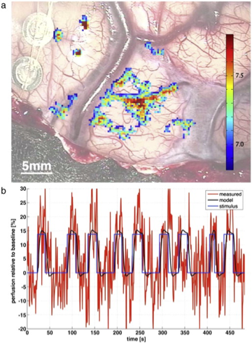
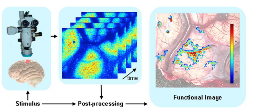
LDF is known for its high temporal resolution (in the range of milliseconds) but its rather poor spatial resolution (in the range of millimeters). Including the baseline reference measurements, intra-operative LDI-acquisition using a single functional task usually spans several minutes (see Figure 1) (5-7). Often, images are presented as color-coded statistical parametric maps, plotted over the video-image of the brain tissue of interest (see Figure 2) (7).
Intra-operative applicability
Like the other optical techniques, LDI is a safe, cheap, portable and non-invasive technique, with intra-operative potential. So far, LDI has been successfully applied in an experimental, intra-operative setting during awake craniotomy surgery. Using a Laser Doppler Imager attached to a conventional microscope, researchers have been able to identify task-based functional regions, using fMRI-style data processing (7). Like all Doppler-based techniques, disentangling tissue motion, global motion artefacts and actual blood flow, are a continuous challenge. What seems to be LDI’s true bottleneck, however, is its poor spatial resolution, combined with its superficial penetration depth, which make intra-operative use for e.g. depth-resolved brain mapping less feasible.
References
- Briers D, Duncan DD, Hirst E, Kirkpatrick SJ, Larsson M, Steenbergen W, et al. Laser speckle contrast imaging: theoretical and practical limitations. J Biomed Opt. 2013;
- Briers JD. Laser Doppler and time-varying speckle: a reconciliation. J Opt Soc Am A. 1996;
- Frerichs KU, Feuerstein GZ. Laser-doppler flowmetry – A review of its application for measuring cerebral and spinal cord blood flow. Molecular and Chemical Neuropathology. 1990.
- Rajan, Vinayakrishnan, Babu Varghese, Ton G. van Leeuwen, and Wiendelt Steenbergen. Review of Methodological Developments in Laser Doppler Flowmetry. Lasers in Medical Science 24, no. 2: 269–83.
- Pouratian N, Toga AW. Optical Imaging Based on Intrinsic Signals. In: Brain Mapping: The Methods. 2002.
- Nielsen AN, Fabricius M, Lauritzen M. Scanning laser-Doppler flowmetry of rat cerebral circulation during cortical spreading depression. J Vasc Res. 2000;
- Raabe A, Van De Ville D, Leutenegger M, Szelényi A, Hattingen E, Gerlach R, et al. Laser Doppler imaging for intraoperative human brain mapping. Neuroimage. 2009;44(4):1284–9.
- Toussay X, Tiberi M, Lacoste B. Laser Doppler Flowmetry to Study the Regulation of Cerebral Blood Flow by G Protein-Coupled Receptors in Rodents. In: Methods in Molecular Biology. 2019.