The following text serves as a temporary placeholder, prior to expert moderation
IT
Moderated by: TBD
Infrared Thermography (IT) or thermoencephalography (TES) is a non-invasive technique which uses infrared radiation (heat) detection as a measure for local neuronal activity (1,2).
Technical Parameters
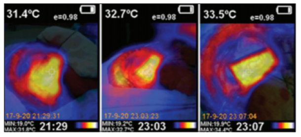
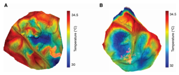
Local increases in neuronal activity are accompanied by increases in local brain tissue temperature – or in other terms – local increases in infrared radiation (1–7). For many years, investigation of this thermoproduction by the nervous system was assessed by placing invasive thermoresistors into the brain (8). Nowadays, this radiation can be detected by a thermoviser camera, which performs line by line scanning of the field of view to eventually create 2D-heatmaps of the brain (see Figure 1 and 2). A single 2D-image can be acquired within several milliseconds and is only a superficial snapshot of the brain cortex, as IT has a depth penetration in the millimeter range. However, a full data acquisition session will take functional tasks of several minutes. The spatial resolution of IT is considered as good, with images presented with <500 µm in pixel size.
Biological Substrate
Neuronal activity is accompanied by local metabolic activity in the brain to satisfy the energetic demands through the process of NVC. This local energy production generates heat which leads to an increase in absolute, local brain tissue temperature concurrent with CBF-changes (6). Although CBF-changes have a large component in the local increase in brain tissue temperature, it is though that the increase is more closely related to actual neuronal activity (6). This is supported by studies such as those by Kiyatkin et al. (9) which indicate that increases in brain tissue temperature occurred prior to and were greater than blood temperature changes evoked by a stimulus.
Intra-operative applicability
ITM has been applied in an intra-operative setting on several occasions, and has proven to be a comparatively cheap, simple and non-invasive technique which can be integrated into conventional surgical microscopes (1,5,10). However, the technique’s sensitivity for functional imaging has been under question on several occasions (4). As alternative applications, studies have mentioned IT as a technique showing potential for surveillance of brain perfusion during vascular neurosurgery (3).
References
- Gorbach AM, Heiss J, Kufta C, Sato S, Fedio P, Kammerer WA, et al. Intraoperative infrared functional imaging of human brain. ANN NEUROL. 2003;54(3):297–309.
- Shevelev IA, Tsicalov EN, Gorbach AM, Budko KP, Sharaev GA. Thermoimaging of the brain. J Neurosci Methods. 1993;
- Rojas EDFR, Ochoa EM, López RL, Díaz LL. Infrared thermography brain mapping surveillance in vascular neurosurgery for anterior communicating artery aneurysm clipping. Surg Neurol Int. 2018;
- Hoffmann N, Radev Y, Koch E, Petersohn U, Steiner G, Kirsch M. Intraoperative mapping of the sensory cortex by time-resolved thermal imaging. Biomed Tech. 2018;
- Shevelev IA. Functional imaging of the brain by infrared radiation (thermoencephaloscopy). Progress in Neurobiology. 1998.
- Suzuki T, Oishi N, Fukuyama H. Simultaneous infrared thermal imaging and laser speckle imaging of brain temperature and cerebral blood flow in rats. J Biomed Opt. 2018;
- Suzuki T, Ooi Y, Seki J. Infrared thermal imaging of rat somatosensory cortex with whisker stimulation. J Appl Physiol. 2012;
- Hayward JN, Baker MA. A comparative study of the role of the cerebral arterial blood in the regulation of brain temperature in five mammals. Brain Res. 1969;
- Kiyatkin EA, Brown PL, Wise RA. Brain temperature fluctuation: A reflection of functional neural activation. Eur J Neurosci. 2002;
- Parrish T, Iorga M. Application of IR thermometry to understanding brain function. In: Quantum Sensing and Nano Electronics and Photonics Xv. Bellingham: Spie-Int Soc Optical Engineering; 2018. (Proceedings of SPIE; vol. 10540).